


Deep, Meaningful AI-Powered Cytology Insights
Insights can make the difference between life and death in cancer detection and treatment. Our advanced AI-powered computational platform helps pathology labs to extract and quantify valuable cellular insights while digitizing 3D cytology samples in minutes.
Schedule Your Personalized Demo
Technology in Action: AIxURO for Bladder Cancer Risk Analysis Using a Urine Sample
- Access ALL fields of view in seconds
- Instantly obtain quantitative scoring across whole samples based on The Paris System (TPS) approach
- Reduce review times
- Securely store data
- Discover clinical insights that can significantly impact patient care
Artificial Intelligence to Reduce Manual Workflow Burdens and Guesswork
Decision Support

Cytopathologists and Cytologists can instantly visualize whole cytology samples and confidently translate quantitative and qualitative data into impactful clinical insights. Precision analysis occurs at both the sample and cell level.
Reproducible and Consistent

Using AIxMed technology, users can reduce time spent manually scanning slides and verifying the work of others. On top of this, any risk of performance variability or unconscious bias is removed so that results are consistent from slide-to-slide and user-to-user.
Smart Automated Scanning

- Whole slide scanning of billions of cells in a 3D volume
- Automatically identify cells of interest
- Calculate precise counts and risk profiles
- Zoom from cell to cell to uncover actionable insights
IT Friendly

Our AI-enabled scanning technology is not just lightning fast, it’s also light on IT infrastructures by using far less power and storage. Digitization of each entire slide is rapid. Files are small to maximize storage space and easily transmit to others.
Distinctly Cytology
In the pathology lab, cytology and histology are quite different. What works for one may not work for another. In fact, many leading FDA-cleared digital pathology systems are designed for histology and are not intended for use with cytology specimens.
 |  |
| Cytology | Histology |
|---|---|
| Cell examination in a fluid matrix | Solid tissue examination |
| 3D volume of cells, typically 30-40 µm deep | 2D flat specimen, typically ~4 µm thick |
| Cells of interest can be anywhere and are difficult to spot in 3D format | Region(s) of interest are typically apparent for closer examination through auto-focus |
| Multiple Z-planes result in very large image file sizes | Single Z-plane results in small image file sizes |
Digital Cytology from AIxMed is the Robust Choice
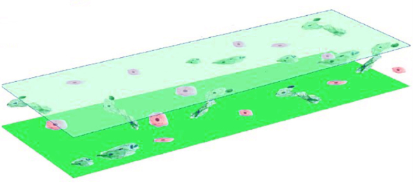
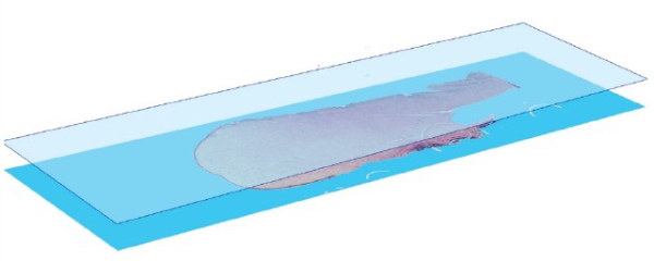
Atypical and suspicious cells could be anywhere in a vast 3D cytology sample slide. If a single focal plane is digitized, cytologists could miss valuable and life-changing opportunities to identify cells of interest.
Using our innovative and proprietary technology, the AIxMed platform rapidly scans multiple Z-planes in the slide’s depth. By doing so, the whole slide is truly scanned. The Z-planes are automatically stacked for an accurate, holistic vision of the patient sample.
Your Insightful Digital Cytology Workflow
Prepare samples using your preferred standard method. Methods may include use of:
- Cytospin™ Cytocentrifuge (Epredia)
- ThinPrep® Pap test (Hologic, Inc.)
- SurePath™ liquid-based Pap test (BD)
Create images using a digital pathology slide scanner.
Upload image files to the appropriate AIxMed computational platform.

Utilize our optional heuristic scanning software to capture images without challenges typically associated with digital cytology:
- Up to 10 slides per hour
- Great cell coverage across multiple Z-planes
- Optimal focal plane search assisted by special light spectrum
- Target cell focusing
- Many image formats accepted
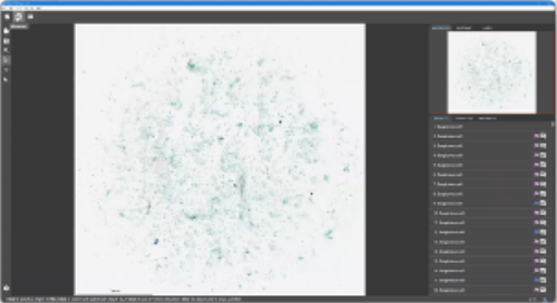
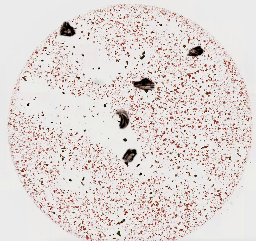
Review quantitative and qualitative data using AIxMed software’s image viewer:
- Large central graphic provides an image overview
- Zoom in to any area for a closer look
- Decision support for urine cytology reporting based on TPS
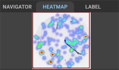
Gain information-rich insights:
- A heat map alerts users to individual cells and other areas of interest
- Individual cells may be sorted according to predictive risk level
- Colors indicate the number of targeted cells and their nucleus-to-cytoplasmic (N:C) ratio
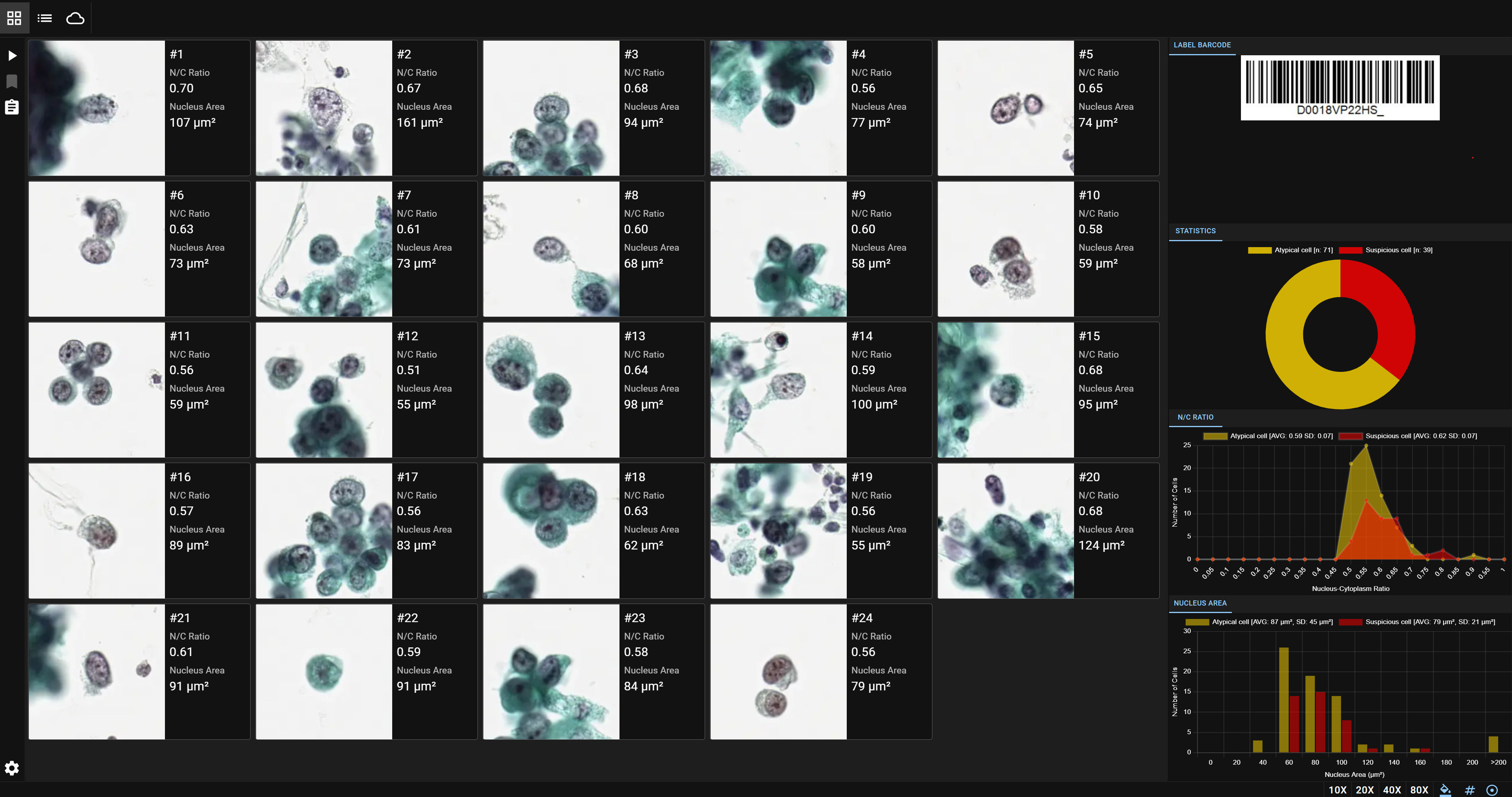
Whole slide quantitative statistics include:
- Total high risk atypia cells
- Total low risk atypia cells
- N:C ratio distribution and mean values
- Nucleus sizes